


0
Views
0
Downloads

0.0
0
0 Likes
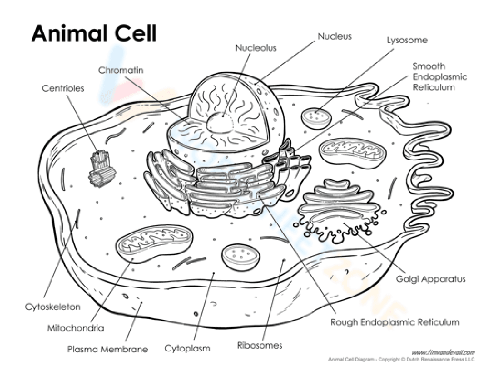
Study an Animal Cell with this Diagram Worksheet
0 Views
0 Downloads
Paste this activity's link or code into your existing LMS (Google Classroom, Canvas, Teams, Schoology, Moodle, etc.).
Students can open and work on the activity right away, with no student login required.
You'll still be able to track student progress and results from your teacher account.
Information
Description
What It Is:
This is a black and white diagram of an animal cell. The worksheet labels the different parts of the cell, including the nucleus, nucleolus, chromatin, ribosomes, smooth endoplasmic reticulum, rough endoplasmic reticulum, Golgi apparatus, lysosomes, mitochondria, centrioles, cytoplasm, cytoskeleton, and plasma membrane. It appears to be designed for labeling or coloring.
Grade Level Suitability:
This worksheet is suitable for grades 6-9. The diagram provides a clear illustration of cell structure and its components, making it appropriate for introducing or reinforcing cell biology concepts at the middle school and early high school levels.
Why Use It:
This worksheet helps students learn the different parts of an animal cell and their locations. It reinforces vocabulary and provides a visual aid for understanding cell structure. It promotes visual learning and memory retention of cell anatomy.
How to Use It:
Students can use this worksheet to label the different parts of the cell using the provided labels. Alternatively, it can be used as a coloring page to help students visualize and remember the different organelles. It can also be used as a review tool to test knowledge of cell structure.
Target Users:
This worksheet is ideal for middle school and early high school students studying biology or life science. It's also useful for teachers looking for a visual aid to supplement their lessons on cell structure. Homeschooling parents can use it to teach cell biology concepts.